
Archaeologists identify a new coelacanth species from a 150-year-old fossil in London. world News

London’s Natural History Museum has discovered a previously unknown species of coelacanth from fossil remains, approximately 150 years old (Macropoma gombesse). The new coelacanth is important because it helps bridge a huge evolutionary gap of about 50 million years for coelacanths, one of the world’s most recognized ‘living fossils’.‘According to the study published by the University of Portsmouth, the fossil was re-evaluated by University of Portsmouth paleontology alumnus Jack L. Norton did this while examining historical collections, which he had already been studying for several years. To explore the internal anatomy of this ancient coelacanth fossil without destroying it, researchers from the University of Portsmouth and the Natural History Museum used advanced X-ray computed tomography (XCT) technology to examine the internal structures of a coelacanth fossil in a way that had never been done before. This discovery emphasizes the enormous amount of scientific potential present in museum collections around the world, and provides further evidence of how modern technological innovations can change the way we look at ancient groups, and show us the complexities of evolutionary processes that have gone unnoticed right under our noses for the past century.
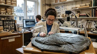
discovery of a New coelacanth species from a 150 year old fossil
As the University of Portsmouth noted, a specimen called Macropoma gombesse was discovered in the Gault Formation of southern England, which dates to the Lower Cretaceous. This fossil has spent over 150 years sitting in plain sight in museum collections, until recently it was discovered that it was a missing link in the Latimeridae family (i.e. modern-day coelacanths). Gombesa is the Comorian name for the modern coelacanth used by local fishermen, from which the name is derived.
Revolutionary discoveries with X-ray computed tomography
The sample was identified using a technique called X-ray computed tomography (XCT), which allows scientists to examine the sample without damaging it, create a 3-dimensional image of the sample, and see the sample’s internal features in high definition. The fossil was then compared directly to similar specimens from different species and found to have enough distinctive structural features to classify it as a new species. It also provided a sample of the evolutionary history of coelacanth.
Revealing internal anatomy with advanced imaging
The researchers used X-ray computed tomography (XCT) to re-analyze the fossil (non-destructive method) through a three-dimensional visualization of the material and internal structures of the specimen; Thus they can perform comparative analysis on this specimen with known lineages to verify any unique physical characteristics of this specimen in relation to other fossils; These results indicate that this fossil represents a new coelacanth species and helps understand its evolutionary history.